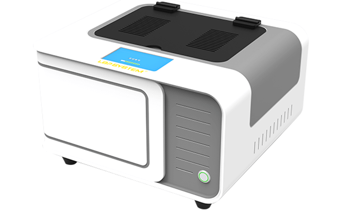

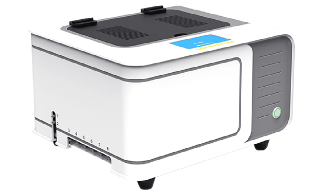
Immunohistochemistry Solutions
From automated staining to biomarker detection, supporting consistent and efficient IHC workflows.
SingPath provides immunohistochemistry solutions covering automated staining systems, IHC antibodies, workflow support, and digital pathology integration.
Designed for clinical pathology and research laboratories, our solutions help standardize IHC processing, improve staining consistency, and support reliable biomarker assessment across a broad range of pathology applications.
Integrated Solutions for the IHC Workflow
Reliable immunohistochemistry depends on both standardized staining and high-quality reagents. SingPath combines automated IHC systems with an expanding antibody portfolio to support laboratories from slide preparation and staining through digital review and biomarker interpretation.
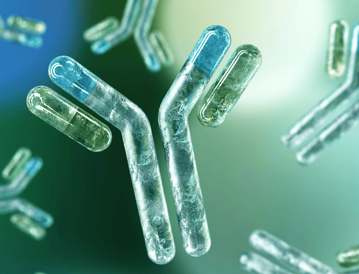
IHC Antibodies
Learn MoreIHC Workflow

Slide Preparation

Baking & Dewaxing

Antigen Retrieval

Primary Antibody Incubation

Detection (Chromogen)

Counterstaining

Digital Scanning

Pathologist Review
01

Slide Preparation
FFPE tissue sections are prepared and mounted onto glass slides.
02

Baking & Dewaxing
Slides undergo preparation to remove paraffin and prepare tissue for staining.
03

Antigen Retrieval
Heat-based antigen retrieval helps expose target epitopes within the tissue.
04

Primary Antibody Incubation
Target-specific antibodies bind to the biomarker of interest.
05

Detection (Chromogen)
The antibody-target interaction is visualized using an appropriate detection system and chromogen.
06

Counterstaining
Tissue morphology is visualized alongside the biomarker signal.
07

Digital Scanning
Stained slides can be converted into high-resolution whole-slide images.
08

Pathologist Review
Biomarker expression and staining patterns are reviewed and interpreted within the clinical context.
IHC Antibody Portfolio
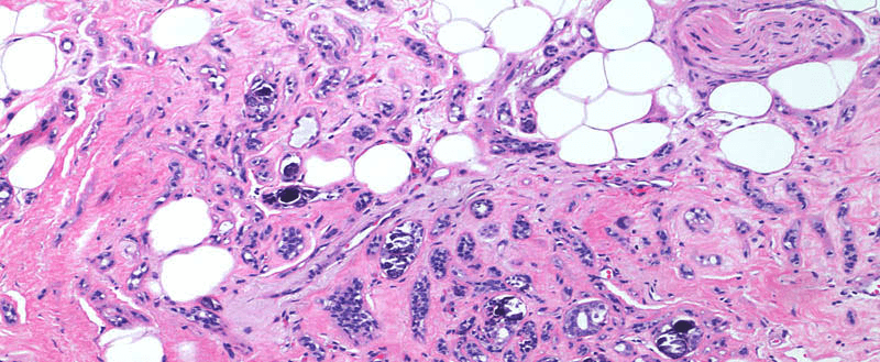
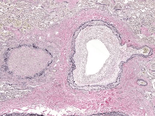
Breast Pathology
Antibodies supporting breast cancer classification, hormone receptor assessment, proliferation analysis, and related biomarker evaluation.
Featured examples:
- ER
- PR
- HER2
- Ki-67
- TRPS1
Thoracic & Lung Pathology
Markers supporting tumor classification, differential diagnosis, and biomarker assessment in lung and thoracic pathology.
Featured examples:
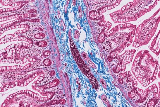
Gastrointestinal Pathology
Markers supporting classification and biomarker evaluation across gastrointestinal malignancies.
Featured examples:
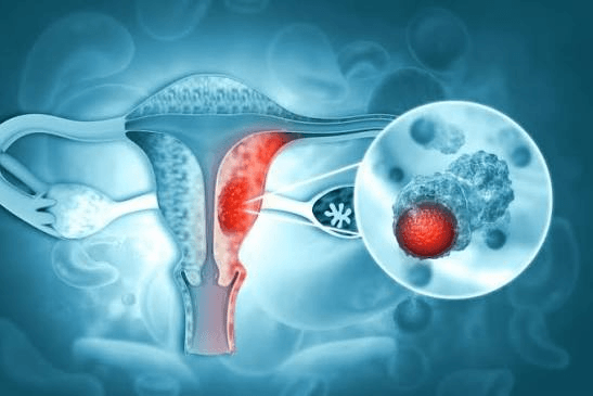
Gynecological Pathology
Antibodies supporting diagnosis and classification of ovarian, cervical, endometrial, and related tumours.
Featured examples:
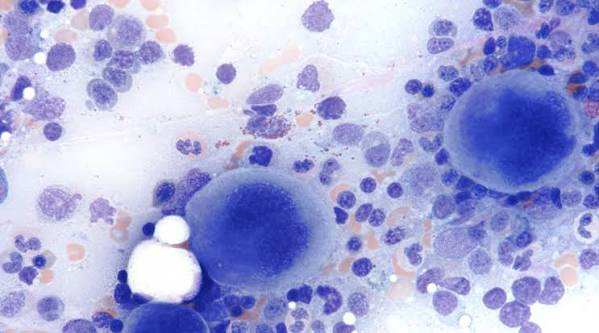
Haematopathology
Markers supporting lymphoid and haematological disease classification and differential diagnosis.
Featured examples:
Precision Oncology Biomarkers
Emerging biomarkers supporting targeted therapy research and precision diagnostic applications.
Featured examples:
- CLDN18.2
- FRα
- c-MET
- PTEN
Beyond Single-Marker Staining
Modern pathology increasingly requires more information from limited tissue samples. SingPath supports workflow development for conventional single-marker IHC as well as multi-marker approaches where different targets can be visualized within the same tissue section.

Single IHC
Routine biomarker detection using one target and one chromogenic signal.

Double Staining
Two biomarkers visualized on the same tissue section using distinct detection signals.

Multiplex Immunofluorescence
Multi-marker fluorescence workflows supporting spatial and tumour microenvironment analysis.