
Fluorescence In Situ Hybridization Solutions
From automated hybridization workflows to targeted probe detection, supporting precise molecular diagnostics.
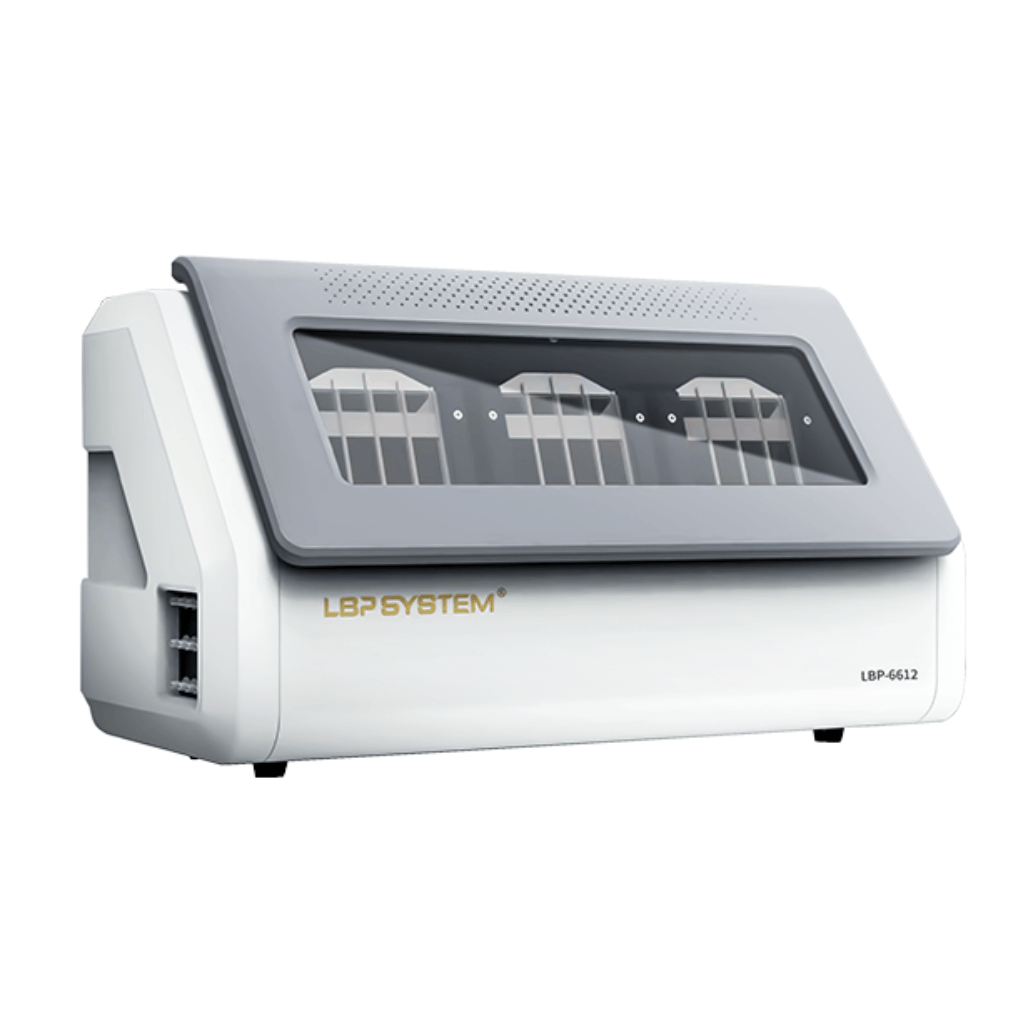
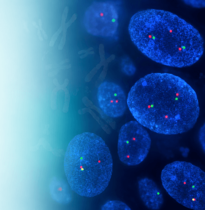
SingPath provides fluorescence in situ hybridization solutions covering automated FISH processing, probe-based detection, workflow support, and digital fluorescence imaging.
Designed for molecular pathology, hematology, oncology, and research laboratories, our FISH solutions help standardize complex hybridization procedures and support the detection of gene rearrangements, amplifications, deletions, and chromosomal abnormalities.
Integrated Solutions for the FISH Testing
Reliable FISH testing depends on both standardized sample processing and high-quality probe reagents. SingPath combines automated FISH instrumentation with a broad probe portfolio to support laboratories across pretreatment, hybridization, washing, signal detection, and result interpretation.
FISH Probes
Learn MoreFISH Workflow

Sample Preparation

Sample Pretreatment

Denaturation

Hybridization

Washing

Counterstaining

Fluorescence Imaging

Signal Interpretation
01
Sample Preparation
Tissue, cytology, or hematological samples are prepared on glass slides according to the test protocol.
02
Pretreatment
Slides undergo appropriate preparation to improve probe accessibility to the target DNA.
03
Denaturation
Target DNA and probe DNA are denatured to allow sequence-specific binding.
04
Hybridization
Fluorescently labelled probes bind to complementary DNA sequences within the sample.
05
Washing
Unbound probe is removed through controlled post-hybridization washing.
06
Counterstaining
Nuclear counterstaining, commonly using DAPI, helps visualize cell nuclei.
07
Fluorescence Imaging
Slides are examined using fluorescence microscopy or digital fluorescence scanning.
08
Signal Interpretation
Signal patterns are assessed to identify relevant genomic abnormalities.
FISH Probe Portfolio
Targeted Probes for Molecular Diagnosis
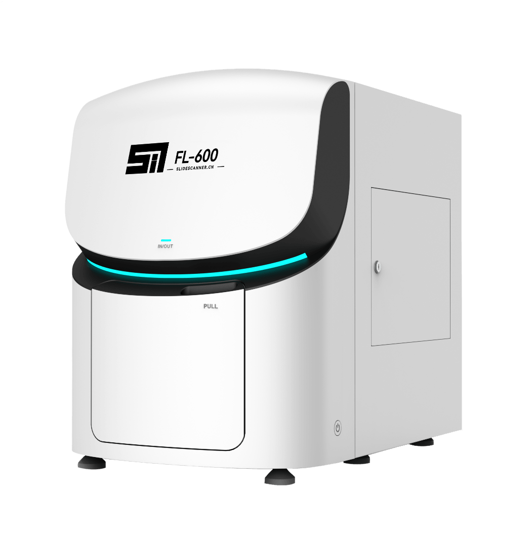
SingPath offers FISH probe solutions supporting the detection of clinically relevant genetic abnormalities across hematology, solid tumors, and precision oncology applications.
The portfolio includes probes for gene rearrangements, translocations, amplifications, deletions, and chromosomal abnormalities.
Hematological Malignancies
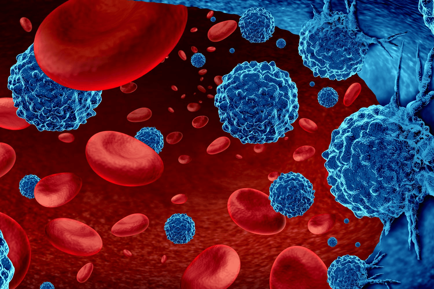
Supporting molecular characterization of leukemia, lymphoma, and myeloid neoplasms
FISH probes can support detection of chromosomal and gene-level abnormalities associated with hematological malignancies:
- Acute Myeloid Leukemia
- Acute Lymphoblastic Leukemia
- Chronic Myeloid Leukemia
- Myelodysplastic Syndromes
- Myeloproliferative Neoplasms
- Lymphoma
- Plasma Cell Disorders


Breast Cancer
Supporting HER2 and related molecular assessment
FISH can be used in selected breast cancer workflows to assess gene amplification and support biomarker evaluation.
Common application
- HER2 amplification assessment
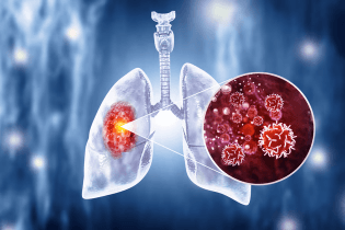
Lung Cancer
Targeted detection of clinically relevant gene rearrangements
FISH probes can support molecular evaluation of selected genomic abnormalities in lung cancer.
Potential applications
- ALK rearrangement
- ROS1 rearrangement
- MET-related abnormalities
- Other clinically relevant targets depending on probe availability
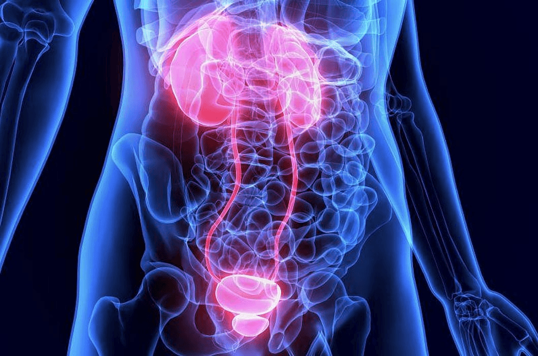
Urological & Genitourinary Tumors
Chromosomal and gene abnormality assessment for selected tumor applications
FISH can support selected diagnostic and research applications in urinary and genitourinary pathology.
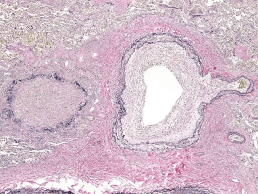
Soft Tissue & Sarcoma
Gene rearrangement detection for tumor classification
FISH is widely used in selected sarcoma and soft tissue tumor workflows where specific gene rearrangements may support diagnosis.
Other Solid Tumors
FISH may support molecular assessment across additional tumor types where amplification, deletion, translocation, or rearrangement status is diagnostically relevant.
Probe Types

Break-Apart Probes
Designed to detect rearrangements involving a specific gene locus without requiring prior knowledge of the fusion partner.
Typical use: Gene rearrangement detection.

Dual-Fusion Probes
Designed to identify specific translocations involving two known genes.
Typical use: Fusion gene detection.

Amplification Probes
Used to assess increases in gene copy number.
Typical use: Gene amplification analysis.

Deletion Probes
Used to identify loss of specific chromosomal regions or gene loci.
Typical use: Deletion analysis.

Enumeration Probes
Used to assess chromosome copy number.
Typical use: Aneuploidy and chromosomal abnormality evaluation.