
 01
Slide Preparation
H&E, IHC, cytology, FISH, or fluorescence slides are prepared using standardized laboratory protocols.
Digital Pathology Solutions
SingPath provides digital pathology solutions covering whole slide scanning, brightfield and fluorescence imaging, digital slide management, remote review, and AI-ready workflows.
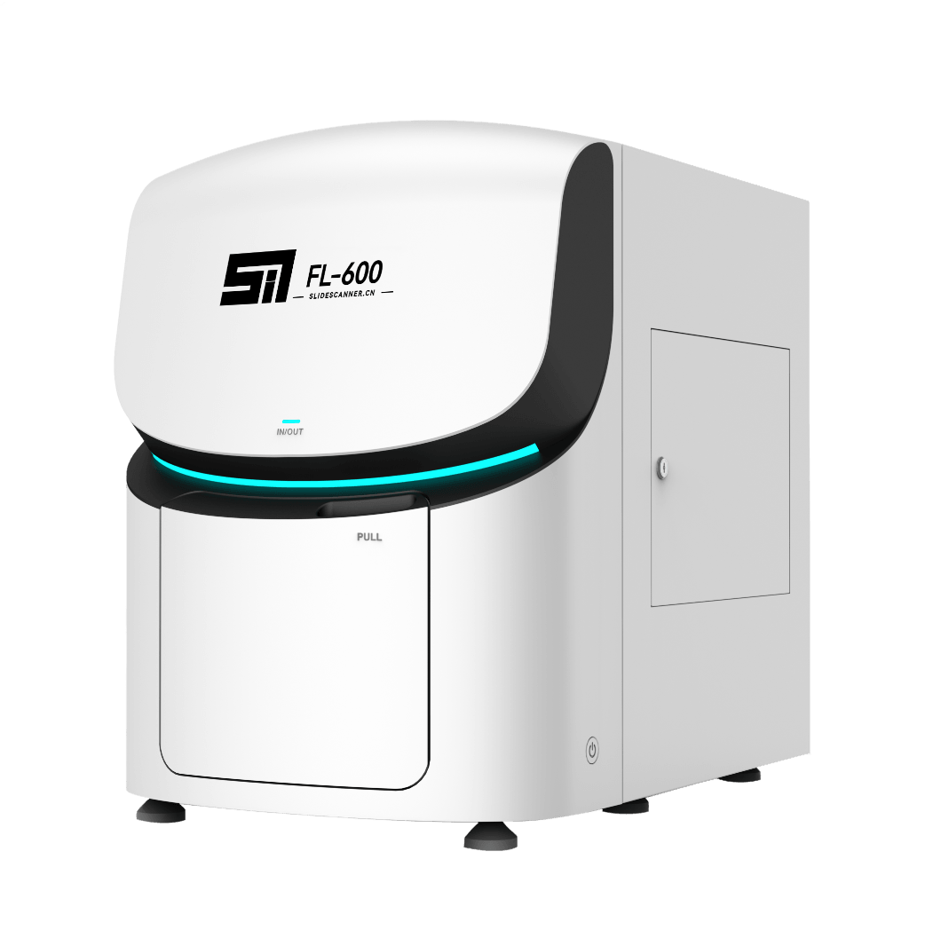
Our scanner portfolio supports laboratories, hospitals, research institutions, and pathology networks in converting glass slides into high-resolution digital images for diagnosis, collaboration, education, research, and image analysis.
Digital pathology extends the traditional microscope workflow by converting pathology slides into high-resolution whole slide images.
Once digitized, slides can be viewed remotely, shared between specialists, archived digitally, integrated with laboratory information systems, and analyzed using image-analysis or AI tools.
SingPath supports this transition with scanning systems for both brightfield pathology and advanced fluorescence applications.
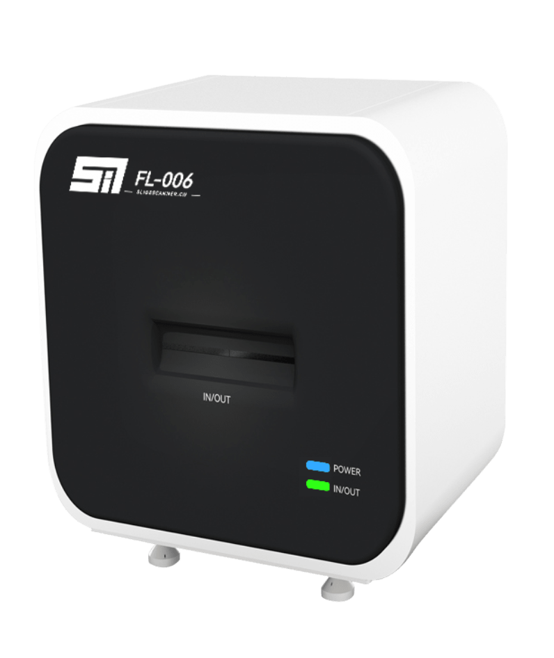
Brightfield digital slide scanners convert conventional stained slides into high-resolution whole slide images suitable for digital review, archiving, consultation, education, and image analysis. Common pathology applications include H&E, IHC, cytology, and special stains.
Typical applications:
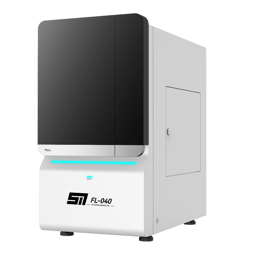
Fluorescence scanners support digital imaging of fluorescence-labelled tissue and cellular samples, enabling multi-channel visualization and analysis for FISH, immunofluorescence, multiplex assays, and research applications.
High-sensitivity fluorescence imaging allows laboratories to capture and preserve signals that traditionally require direct fluorescence microscopy.
Typical applications:
Slide Preparation

Slide Loading

Identification

Whole Slide Scanning

Image Processing

Digital Review

Data / AI Analysis
01H&E, IHC, cytology, FISH, or fluorescence slides are prepared using standardized laboratory protocols.
02Slides are loaded into the scanner individually or in batches according to scanner capacity.
03Slide labels and case information can be captured to support specimen identification and digital workflow management.
04The scanner identifies the tissue region and captures high-resolution images across the slide.
05Captured images are assembled into a navigable digital whole slide image.
06Pathologists can navigate, zoom, annotate, compare, and review slides digitally.
07Digital images can be used for image analysis, quantitative assessment, or AI-assisted workflows.

Capture detailed whole slide images suitable for pathology review and image-based analysis.

Automatically identify relevant tissue regions to support efficient scanning workflows.

Maintain image clarity across uneven tissue sections and varying slide surfaces.

Support slide identification and digital case association through label and barcode recognition.

Capture multiple focal planes for samples requiring depth information.

Support automated high-volume processing for laboratories with demanding slide workflows.

Navigate high-resolution pathology images with smooth zoom and pan functionality.

Organize digital slides by patient, case, specimen, or laboratory workflow.

Mark regions of interest, add annotations, and perform image measurements.

Compare different stains or related slides within the same case.

Maintain searchable digital slide libraries for retrospective review and teaching.

Support integration with laboratory information systems according to project requirements.